
Joint preservation procedures aim to maintain and restore the function of joints without resorting to joint replacement. These procedures are particularly beneficial for younger, active patients who suffer from joint pain due to injury, early arthritis, or other joint conditions. Here’s an overview of common joint preservation techniques:
Arthroscopy:
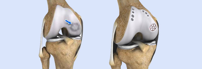
Cartilage Restoration:
Ligament Reconstruction:
Osteotomy:
Arthroscopy:
Rotator Cuff Repair:
Labral Repair:
Biceps Tendon Repair or Tenodesis:
Arthroscopy:
Periacetabular Osteotomy (PAO):
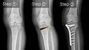
High tibial osteotomy (HTO) is a surgical procedure that realigns the knee joint by cutting and repositioning the tibia (shin bone). The procedure is often used to treat medial knee arthrosis, which can cause a bowlegged alignment that puts too much stress on the knee's inner compartment. HTO can also be used to correct varus or valgus alignment, knee instability, post-traumatic arthritis, and knee pain.
During the procedure, a wedge of bone graft or synthetic bone is placed on the medial side of the tibia and secured with screws and a plate, or a wedge of bone is removed from the outer side. HTO is usually recommended as a last resort after other non-surgical treatments have been tried.
Studies have shown that HTO can produce good long-term results if the patient is selected correctly and the surgery is performed precisely. For example, one study found that patients who had HTO were able to return to sports and other activities more quickly than those who had UKA, and also had better sports-related functional scores two years after the procedure. Another study found that HTO resulted in a better range of motion after surgery than UKA.
However, HTO can also have complications, including infection, loss of correction, and nonunion. There's also a risk of lateral hinge fracture if the HTO is medially based, and a risk of peroneal nerve injury if it's laterally based. About 15% of patients may need to have the procedure redone, with hardware removal being the most common procedure.
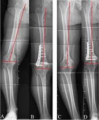
A distal femoral osteotomy (DFO) is a surgical procedure that involves cutting and straightening the femur (thigh bone) to correct knee alignment. The goal of the procedure is to realign the knee so that the weight-bearing axis passes through the center of the knee or slightly into the inside compartment. This can help prevent excessive loading and degeneration of one side of the knee joint.
DFO is often used to treat patients with knock knees, also known as valgus knees, where the knee's mechanical axis passes laterally of the knee joint center. It can also be used to treat patients with arthritis on the outside of the knee.
There are two types of DFO: Medial closing osteotomy (MCDFO) and Lateral opening distal femoral valgus osteotomy (LODFO).
The procedure involves creating a surgical fracture at the end of the femur and repositioning the bones so they are secured in the proper alignment. After the procedure, patients typically spend at least one night in the hospital and wear a range of motion brace for at least two months while the bone heals. They may also be prescribed antibiotics for at least six days and blood thinners, and they may only be allowed to partially bear weight for two months.
If you would like to have additional information contact Dr. Vipul Shet, serving communities and people from all walks of live.